Oral Myiasis: When Reality Mimics Horror
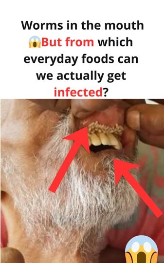
It may sound like a plot from a chilling horror film, but oral myiasis is a documented and serious medical reality. This condition occurs when fly larvae, commonly known as maggots, infest, grow, and survive within the human oral cavity.
While statistically rare on a global scale, cases are consistently reported in regions where sanitation is poor, fly populations are high, and access to professional dental or medical care is limited. For most, the idea is unthinkable, yet oral myiasis serves as a grim reminder of the human body’s vulnerability when hygiene is compromised or environmental conditions allow such an infestation to take hold.
Understanding Oral Myiasis
In medical terms, myiasis refers to a parasitic infestation caused by the larvae of specific fly species. these larvae sustain themselves by feeding on living or necrotic (dead) tissue, bodily fluids, or even ingested food.
When this parasitic process localizes inside the mouth, it is classified as oral myiasis. The infestation can target various areas, including:
- The gums and palate
- The inner lining of the cheeks
- The tongue and lips
In advanced or severe cases, the presence of these larvae can lead to extensive soft tissue damage, compromise vital oral functions like speaking and eating, and potentially spread to adjacent facial structures.

Oral myiasis can affect individuals of any age, from children to adults. However, the risk is significantly higher among the elderly, the disabled, and those with compromised immune systems. Individuals who struggle with self-care—such as those suffering from paralysis, neurological disorders, or chronic alcoholism—are particularly susceptible.
The primary catalyst for this condition is often a combination of poor oral hygiene and advanced dental disease. Flies are drawn to moist, exposed, or necrotic (dead) tissue, which provides the perfect nutrient-rich environment for their larvae to flourish.
The Development of an Infestation
The process begins when opportunistic fly species deposit their eggs into vulnerable areas of the mouth. This often occurs in individuals who are unable to maintain oral cleanliness or those who sleep with their mouths partially open, providing easy access for the insects.
Several fly species are commonly associated with this condition, including:
- Chrysomya bezziana: The Old World screwworm fly.
- Cochliomyia hominivorax: The New World screwworm fly.
- Lucilia sericata: The green bottle fly.
- Musca domestica: The common housefly (in rarer instances).
Once the eggs are laid, they hatch into larvae within hours or a few days. These larvae immediately burrow into the soft tissues of the mouth, feeding on food debris and tissue. This feeding process causes progressive destruction of the oral structure and leads to the distressing symptoms characteristic of the infestation.
Symptoms and Early Warning Signs
Early detection is vital to preventing extensive tissue damage. Oral myiasis typically manifests through a series of progressive symptoms, including:
- A “Crawling” Sensation: A distinct feeling of movement beneath the tissues of the mouth.
- Physical Inflammation: Significant swelling, redness, and irritation in the affected area.
- Functional Difficulty: Pain or discomfort while chewing, swallowing, or speaking.
- Increased Salivation: Excessive drooling or a constant bad taste/foul odor.
- Visible Larvae: In advanced stages, tiny white or yellowish moving larvae may be visible between the gums, on the tongue, or within the inner cheeks.

If left untreated, an infestation can escalate with terrifying speed. The larvae’s constant feeding leads to necrosis (tissue death) and creates an opening for secondary bacterial infections. In extreme and rare circumstances, the larvae can migrate beyond the mouth, invading surrounding facial structures and deep tissues, which necessitates emergency surgical intervention to prevent permanent disfigurement or systemic complications.
Debunking Common Misconceptions
There is a widespread myth that oral myiasis is caused by eating contaminated or “spoiled” food. However, medical evidence shows that direct infection through ingestion is extremely rare.
The primary mode of transmission is direct contact. This occurs when flies land on or near the mouth, particularly while a person is sleeping or if they have limited physical mobility to shoo the insects away. Several factors create the “perfect storm” for this to happen:
- Untreated Dental Disease: Open sores, ulcers, or decaying gum tissue act as a magnet for flies.
- Mouth-Breathing: Chronic mouth-breathing or the inability to close the mouth fully provides easy entry for pests.
- Systemic Vulnerability: Conditions like diabetes, HIV, or severe immune suppression weaken the body’s natural defenses.
It is crucial to understand that oral myiasis isn’t simply a result of “bad luck” or eating poorly; it is the result of specific environmental and physiological vulnerabilities intersecting.
Real-World Impact: Case Studies
Documented medical reports from around the globe highlight the devastating nature of this condition. In various regions of India, Brazil, and Africa—where fly density is high and specialized dental care is often out of reach—cases frequently involve the elderly or the bedridden.
- Hospital Reports: One recorded case described a patient arriving with dozens of larvae nested within the oral cavity, resulting in severe hemorrhaging and necrotic gum tissue.
- Vulnerability in Care: Another incident involved a bedridden elderly patient whose inability to maintain basic oral hygiene, combined with constant mouth-breathing, led to a deep-seated infestation.
In almost all documented cases, the removal of the larvae is a delicate process that requires medical professionals to work under local or general anesthesia, followed by aggressive antibiotic treatment to manage secondary infections.
Clinical Diagnosis
Diagnosis is primarily clinical, meaning it is identified through direct observation. Medical professionals look for specific indicators, such as:
- Larval Movement: The presence of live, moving larvae within the folds of the oral cavity.
- Tissue Presentation: Highly inflamed, foul-smelling, or necrotic tissue surrounding a central site of infestation.
Secondary bacterial infections or foul-smelling exudate
Imaging studies such as MRI or CT scans are occasionally used in severe cases to assess tissue involvement and ensure that deeper structures are not affected.
Laboratory tests may include parasite identification to determine the species of fly involved, which can inform treatment and prevention strategies.
Treatment
Treatment of oral myiasis is urgent and methodical, requiring both mechanical removal of larvae and medical management. The primary steps include:
Manual Extraction:
Each larva is carefully removed using forceps or tweezers.
Local anesthesia is typically used to reduce pain and ensure patient compliance.
Pharmacological Intervention:
Ivermectin, an antiparasitic medication, may be administered to kill larvae that are embedded deeper in tissue and not visible.
Antibiotics are often prescribed to control or prevent secondary bacterial infections.
Wound Care and Rehabilitation:
Affected tissues are disinfected and cleaned regularly.
In severe cases, surgical reconstruction may be necessary to restore gum tissue, inner cheek integrity, or even lips if extensive necrosis has occurred.
Dental Follow-Up:
Patients often require long-term dental care to restore oral function and aesthetics.
Prognosis
With timely and comprehensive treatment, most patients recover fully, though severe cases may require multiple interventions.
Early recognition dramatically improves outcomes and reduces the risk of tissue loss, systemic infection, or recurrence.
Prevention
Fortunately, oral myiasis is largely preventable. Key preventive measures include:
Good oral hygiene: Brushing teeth twice daily, flossing, treating gum disease, and regular dental check-ups.
Maintaining a clean living environment: Proper disposal of waste, minimizing fly exposure, and keeping sleeping areas free of insects.
Protecting oral wounds: Covering sores, ulcers, or damaged tissue to prevent fly access.
Monitoring at-risk individuals: Elderly, disabled, or unconscious patients should have daily oral care performed by caregivers.
Even simple precautions, such as using mosquito nets or window screens, can reduce the likelihood of flies contacting oral tissue.
Public Awareness
While oral myiasis is alarming, it is important to remember that it is extremely rare in populations with normal hygiene and access to healthcare.
Educating healthcare providers, caregivers, and vulnerable populations is critical to early detection and rapid intervention.
Recognizing the early signs — crawling sensations, excessive salivation, swelling, or foul odor — allows prompt medical action, preventing severe outcomes.
Conclusion
Oral myiasis represents a unique intersection of biology, hygiene, and human vulnerability.
It is a stark reminder that even in a modern, medically advanced world, parasitic organisms can exploit weaknesses in human tissue under certain conditions.
While the thought of living larvae inside the mouth is horrifying, understanding the causes, symptoms, treatment, and preventive measures can reduce fear and empower individuals to protect themselves.
The human body has remarkable defenses, but oral myiasis is an unsettling example of nature’s persistence and opportunism. Awareness, hygiene, and timely medical care remain the most effective tools for prevention and recovery.